

Project Description
RETINA
Home > Our Ophthalmologist > Services > Retina
The retina is a very important structure in the eye, composed mainly of nerve fibers which allow the images we see to be transmitted to the brain. There are some common systemic conditions, such as diabetes which can affect the retina and the macula resulting in poor vision for the patient. Other conditions such as age-related macular degeneration may also result in poor vision for the patient.
Diabetic Eye Screening
As diabetes is a chronic condition, patients may develop diabetic eye disease in the long run. Patients who are not compliant to keep their blood sugar levels low may be at a higher risk of developing diabetic eye disease as well. Diabetic eye disease can present itself through a few ways. They include diabetic macular edema, diabetic retinopathy, vitreous haemorrhage, neovascular glaucoma and cataract.
Therefore, patients who have already been diagnosed with diabetes are highly recommended to have their eyes screened.
Diabetic Eye Diseases
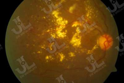
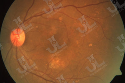
Diabetic retinopathy is caused by retinal ischaemia where there is lack of blood supply to the retina which results in formation of weak new blood vessels and leakage of blood vessels. This will result in bleeding of the eye and swelling of the retina.
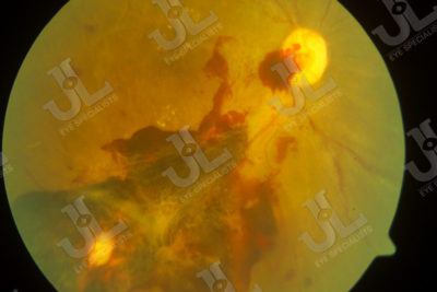
Vitreous haemorrhage is when the leaked blood in the retina flows to the clear gel like substance (vitreous) of the eye.
Diabetic macular edema or diabetic maculopathy is when there are leakages of blood vessels and swelling of the macula. This is the most common cause of poor vision in a patient as the macular is part of the retina that gives us good vision.
Neovascular glaucoma develops in eyes with late stages of diabetic eye disease. The eye responds to the lack of blood supply to the eye by producing abnormal new blood vessels at the front part of the eye, typically seen on the iris and obstructing the fluid drainage channel of the eye. This would cause complications such as pressure build up in the eye, resulting in a severe optic nerve damage.
Diabetes can lead to cataract formation in the eye as uncontrolled diabetes causes sugar levels to increase in the aqueous fluid and affecting the lens. This would then lead to progressive blurring of vision over time.
Long-term diabetes and poorly controlled diabetes can cause reduced blood flow to the eye. This may result in the ischaemia of the optic nerve.
Management of Diabetic Retinopathy
The approach to the management of diabetic retinopathy is to first detect it. During the eye screening, Dr Jimmy Lim would be able to perform tests to detect presence of diabetic retinopathy and macular edema even before the patient begins to notice any symptoms in the vision. Therefore, it is important for patients to undergo eye screening at least once a year.
For patients who have been diagnosed with retinopathy or maculopathy, it is important to have a tighter control of their diabetes.
Patients will need various types of lasers to decrease the risk of severe vision loss and vitreous hemorrhage, and treatment includes the use of intravitreal anti-VEGF injection to decrease the swelling of the macular and improve the patient’s vision.
Even after treatment, it is essential for patients to keep their diabetes in control and have regular follow-ups with the eye specialist to monitor the progression of the disease.
Age-Related Macular Degeneration
Age-Related Macular Degeneration (AMD) mainly affect older patients, with them complaining of distortion or loss of central vision. This is because of degeneration to the macular (center part of the retina) may result in loss of good central vision.
Types of Age-Related Macular Degeneration
Age-Related Macular Degeneration can be classified into dry and wet AMD.
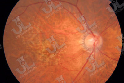
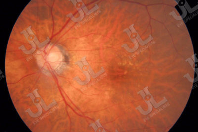
Dry AMD usually have no visual symptoms to mild visual symptoms and it is detected upon examination by an eye doctor. It is detected with drusen which can be seen over the macular and there may be a thinning (atrophy) of the macular area. Dry AMD may progress to wet AMD if it goes undetected.
Wet (Exudative) AMD have visual symptoms that range from mild to severe loss of central vision. There may be swelling of the macular and even bleeding at the macular area. The nerve fibre layer of the retina can be detached and there may also be vitreous haemorrhage.
Polypoidal Choroidal Vasculopathy (PCV) also presents with signs and symptoms similar to wet AMD. Visual symptoms can range from mild to severe loss of central vision. There may be swelling of the macular and bleeding at the macular region. Detachment of the nerve fibre layer and vitreous haemorrhage can also be seen.
Management for Age-Related Macular Degeneration
Management for dry AMD is to prevent or reduce progression of the condition by taking more fresh fruits and vegetables. Diet should be complemented with nutritional supplements of lutein and zeaxanthin. A Home Amsler Chart is use to monitor for any changes to visual symptoms. Patients should also avoid or quit smoking cigarettes or tobacco.
On top of the management steps for dry AMD, wet AMD will also require intravitreal injections of anti-VEGF (anti-Vascular Endothelial Growth Factor) to prevent further swelling or bleeding of the macular area. Laser treatment may also be indicated to treat wet AMD.
Management of PCV is usually by intravitreal injection of anti-VEGF and through laser treatment with Photodynamic therapy.
Much research has been done on the use of intravitreal anti-VEGF injection for the treatment of wet AMD, and its use has successfully improved the vision of patients with wet AMD.

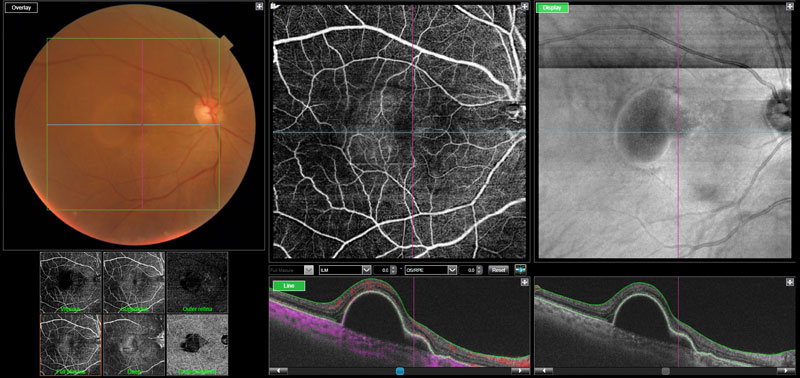
Image 4.1 The swept-source OCT Angiography report shows a degeneration of the macular with pigment epithelium detachment.
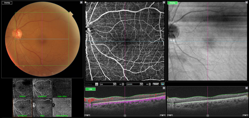
Image 4.2 The swept-source OCT Angiography shows a healthy macula with a regular and smooth retina layer with no detachment of the pigment epithelium layer.
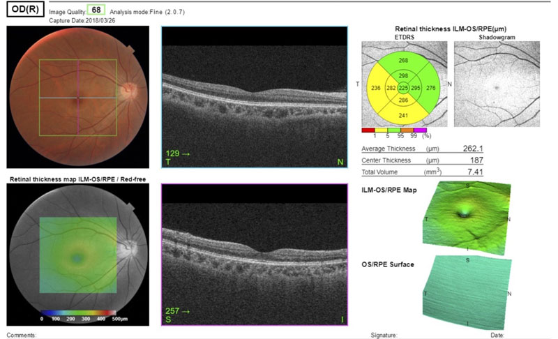
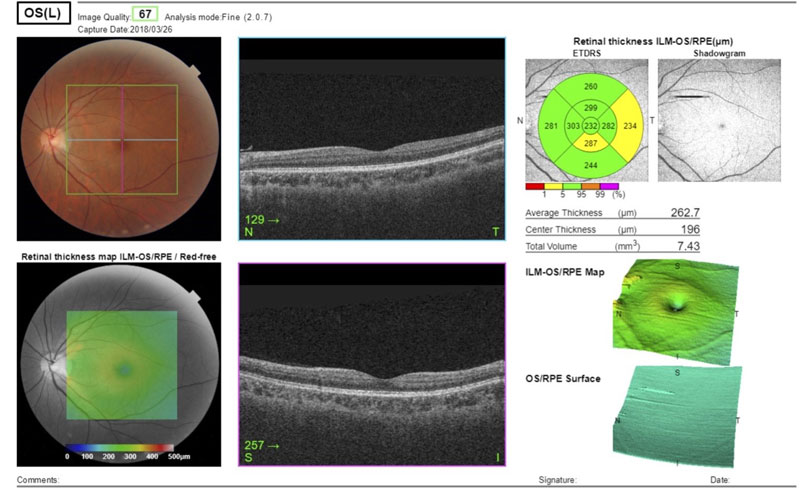
Image 4.3 & 4.4 Show an OCT scan of a healthy macular. The architecture of the retina is smooth and without any irregularities, and the pigment epithelium layer is not detached from the choroidal layer below.
Advanced Technologies for Diagnostics and Management of Age-Related Macular Degeneration and Diabetic Retinopathy
DRI OCT Triton
Optical Coherence Tomography (OCT) is a non-invasive diagnostic imaging test which uses light waves to take cross-section or 3 dimensional pictures of a person’s retina. OCT allows each of the retinal layers to be distinctively seen so that they can be differentiated and have their thickness measured.
Comparing the thickness of a patient’s retina to a normal heathy retinal layer can help doctors determine whether retinal disease exist before the patient even complains of any symptoms. Using an OCT also allows for the early treatment in patients and improves the success of these treatments, especially in wet macular degeneration, where the eye disease progresses quickly.
The DRI OCT Triton at JL Eye Specialists performs a combined anterior and posterior swept-source OCT, providing a significant improvement over the conventional OCT. Due to the optimized long wavelength scanning light (1,050nm), there is better penetration of the deeper layers of the eye. This scanning light also penetrates better through cataracts, hemorrhages, blood vessels and sclera.
The DRI OCT Triton also bring more scans for a single B-scan image, and more informative image supports efficiency and quality of diagnosis. The high penetration of the swept-source light can easily and clearly visualize deep layers in the eye, such as choroid and sclera. A further benefit of swept-source is that it can clearly visualize both the vitreous and choroid in a single scan, that are uniformly clear and noise-free. This eliminates the need for time consuming vitreous/choroidal combination scans.
Swept-Source OCT Angiography
The DRI OCT Triton combines high quality OCT angiography with a swept-source OCT.
This system is built on the clinically proven DRI OCT Triton platform, powered by OCTARA, an algorithm that provides highly sensitive angiographic detection. The choroid and even deeper retinal layers are imaged exceptionally with this feature. It uncovers low vascular flow with high sensitivity. In addition, the 1μm wavelength feature allows OCT imaging even for patients with media opacities.
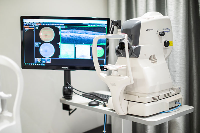
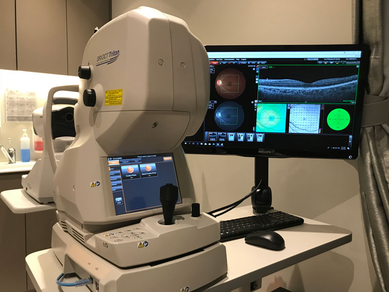
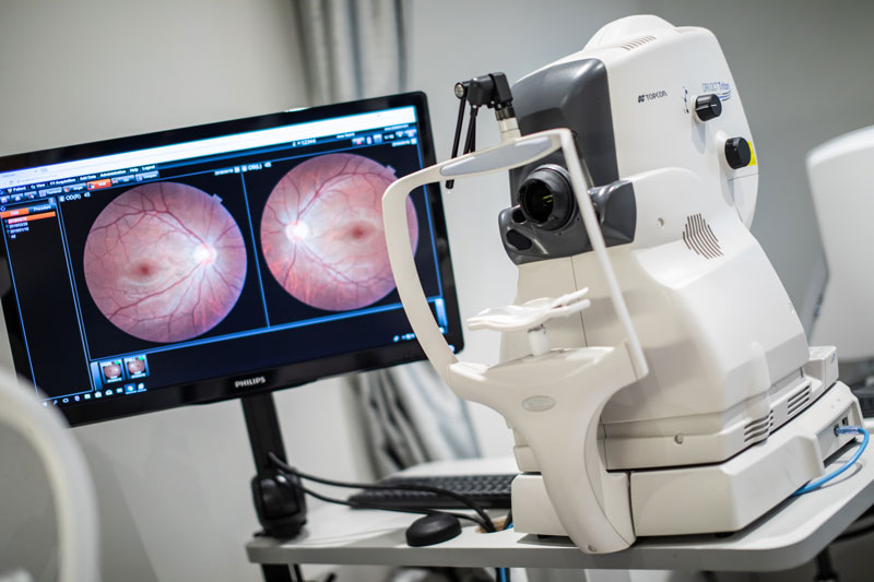
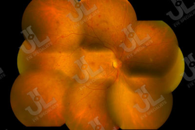
Image 4.5 shows a fundus image of a patient taken using the DRI OCT Triton machine at JL Eye Specialists. The photo shows a 30 degree field of view of the central retina with the optic nerve in view.
Book An Appointment With Us
We understand that each patient comes with their unique concerns. We customize our approach to ensure that we provide each patient with the appropriate treatment to address their needs.