
Project Description
GLAUCOMA
Home > Our Ophthalmologist > Services > Glaucoma
Glaucoma is a silent thief of eyesight. The disease progressively damages the optic nerve of the eye without any signs or symptoms till very late in the process when patients realize a narrowing of the visual field or blurring of vision.
Several studies have shown that eye pressure is a major risk factor for optic nerve damage. The excessive build up of pressure in the eye occurs because of an imbalance between fluid production and its drainage.
There are different types of glaucoma:
- Open-angle glaucoma
- Closed-angle glaucoma
- Low or normal tension glaucoma
Open-Angle Glaucoma
This is the most common type of glaucoma and it affects the elderly and some who are middle-aged. It happens gradually where the eye does not drain fluid as well as it should, resulting in an increase in the eye pressure that may damage the optic nerve. The glaucoma progresses slowly and painlessly, so the patient may not feel any symptoms and slight loss of vision may go unnoticed.
Closed-Angle Glaucoma
In acute closed-angle glaucoma, the patient can suddenly experience pain and rapid vision loss. Fortunately, the symptoms of pain and discomfort make the patient seek medical help, resulting in prompt treatment, which may prevent further damage from occurring.
In chronic closed-angle glaucoma, symptoms that patients may experience are similar to open-angle glaucoma in that it may progress slowly and painlessly, and patients may not feel any symptoms and slight loss of vision may go unnoticed.
Low or Normal Tension Glaucoma
Even though eye pressure is normal, optic nerve damage still occurs. It might be due to reduced blood supply to the optic nerve.
All three types of glaucoma have the common end point of damage to the optic nerve and visual field loss. Thus, screening for glaucoma is very important especially for patients in the following groups:
- Family history of glaucoma
- History of high eye pressure
- History of injury to the eye
- History of inflammation of the eye
- Older age group
Glaucoma Screening
The tests that are performed to screen for glaucoma are:
- Eye pressure checks
- Eye and optic nerve examination
- Visual field studies
- Anatomical studies of the optic nerve compared to normal population (looking at the structure of the optic nerve)


Management of Glaucoma
Glaucoma can be treated with eye drops, laser treatment or surgery, or a combination of these methods.
Anti-glaucoma eye drops are often the first choice over surgery and can be very effective at controlling the intraocular pressure (IOP). Some patients may be prescribed with more than one type of eye drop to achieve the best IOP control.
Laser treatment and surgical options are available for cases where glaucoma damage is more severe with poor or difficult eye pressure control.
Several types of laser treatments are available to either reduce the production of fluid in the eye or improve the drainage outflow of the eye.
There are also innovative surgical options where surgery is performed to improve drainage outflow of the eye. Glaucoma drainage devices that may also be implanted into the eye for the same purpose.
The goal of any treatment is to prevent loss of vision, as vision loss from glaucoma is irreversible. Fortunately, glaucoma can be managed if detected early, and with medical and/or surgical treatment, most people with glaucoma will not lose their sight.
Advanced Technologies for Glaucoma Diagnostics and Management
Octopus 900
The Octopus 900 is a highly versatile machine used to measure an individual’s central visual field. Depending on the patient’s eye condition and symptoms, the doctor can order for specific type of tests to check for any abnormalities or loss of visual field. A full threshold testing of the visual field can be done in just 2-4 minutes, which is much faster comparing to other visual field machines, thus reducing patient fatigue while going through the test. It also allows the doctors to easily interpret the results, allowing for timely intervention should there be any visual field loss.
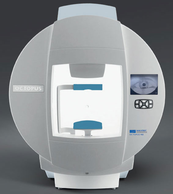
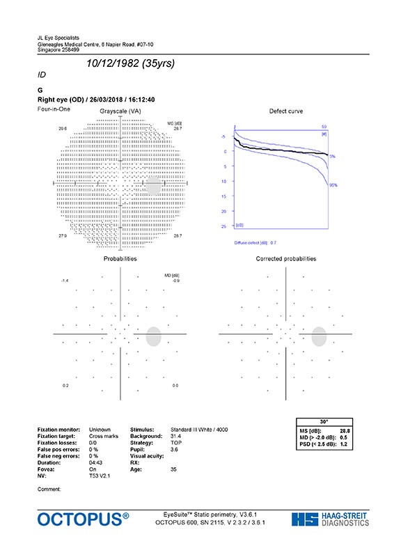
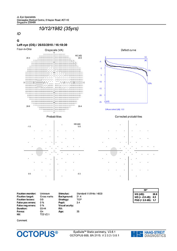
Image 3.1 and 3.2 The normal visual field reports of the right and left eye of the patient. The results fall within the normative values of the population as indicated by the chart for both eyes even though there may be slight differences in the visual field patterns between the two eyes.
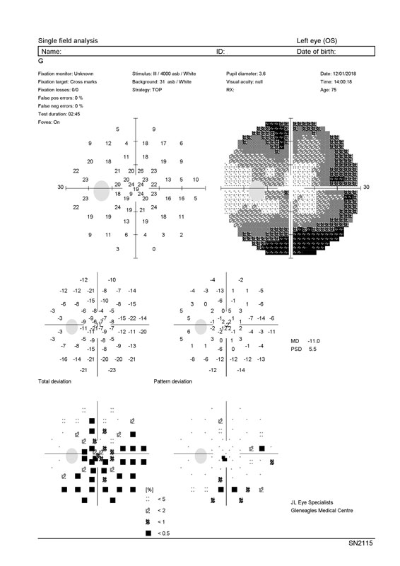
Image 3.3 shows a diffused visual loss from the left eye of a patient with glaucoma. Damage to the optic nerve fibres reduce their sensitivity to the stimulus, thus it is picked up by the test and reflected in the report.

Image 3.4 presents with a superior visual field loss on the left eye, indicating damage to the inferior segment of the optic nerve fibres. With specific patterns on the report and symptoms from the patient, the doctor can diagnose if the visual field loss is due to glaucoma, its progression, or any other underlying conditions.
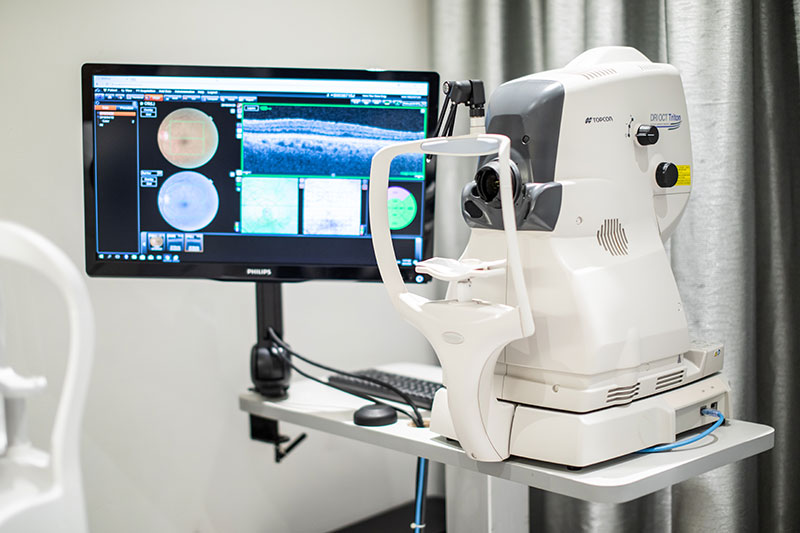
DRI OCT Triton
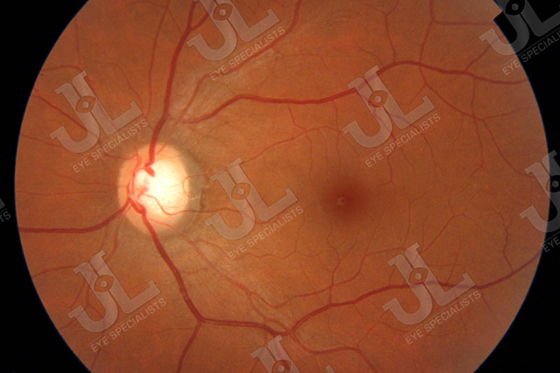
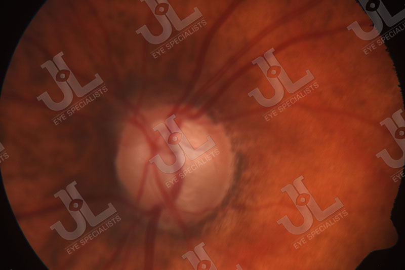
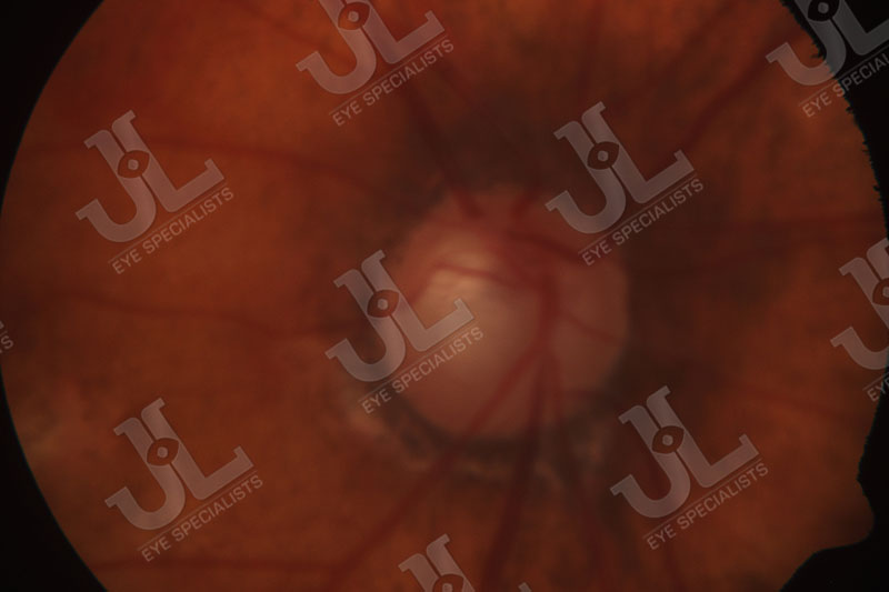
The Triton Optical Coherence Tomography (OCT) takes detailed images of the retinal nerve fibre layer (RNFL) at the optic nerve. It captures precise three-dimension images and it can clearly portray the extensiveness or the progression of damage to the nerve. The system is also able to display the results, comparing the thickness of the patient’s RNFL to that of a population matched to the patient’s race and age group. Thus indicating if the changes are within the normal or abnormal limits.
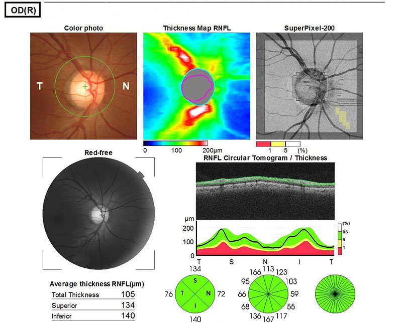
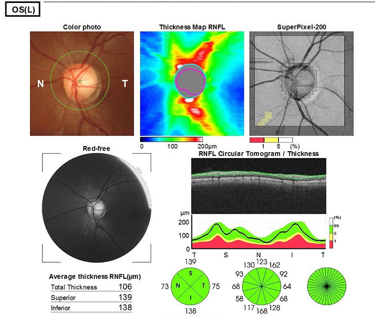
Images 3.5 and 3.6 are normal reports from the OCT which shows that the patient’s RNFL thickness are within normal limits when compared to a population matched to the patient’s age and race group.
Book An Appointment With Us
We understand that each patient comes with their unique concerns. We customize our approach to ensure that we provide each patient with the appropriate treatment to address their needs.