

Project Description
CORNEA
Home > Our Ophthalmologist > Services > Cornea
Cornea and Ocular Surface Diseases
The cornea is a clear structure that is found in the front of the eyeball. It serves as a very important refractive surface of the eye. The cornea’s regular surface and its clarity allows light to enter our eyes, giving us good vision.
Therefore, any conditions causing irregularity, scarring, swelling and opacities of the cornea will result in poor vision for the patient.
Our Medical Director Dr Jimmy Lim is a cornea specialist with many years of experience in diagnosing and managing cornea and ocular surface diseases.
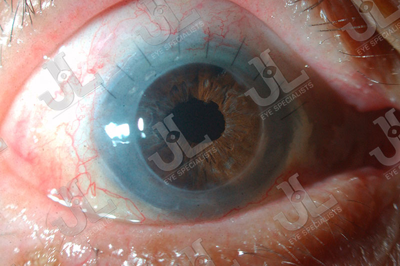
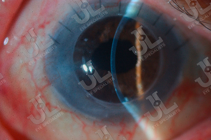
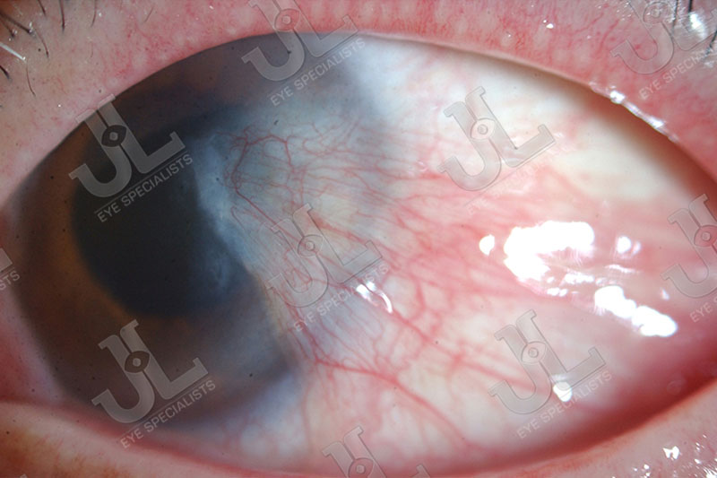
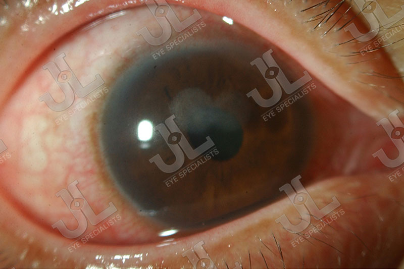
Cornea Decompensation or Scarring
Cornea decompensation or scarring is the failure of the cornea to maintain its clarity. People with this condition experience poor vision and discomfort in the eye, and have a high risk of cornea infection.
Cornea decompensation or scarring is usually caused by a previous cornea infection or trauma to the cornea. The condition could also be brought about by cornea dystrophy (eg. Fuch’s Endothelial Dystrophy) and cornea swelling or edema.
Treatment of Cornea Decompensation or Scarring
Medication has very limited effect in treating cornea swelling. Laser treatment such as PhotoTherapeutic Keratectomy (PTK) would be able to clear some of the superficial scarring.
Cornea Transplant
Cornea transplant is a surgical procedure where a damaged or diseased cornea is replaced by a donated corneal tissue (the graft) taken from a recently deceased individual.
When the entire or full thickness of the cornea is replaced, it is known as penetrating keratoplasty. During the procedure, a cornea button that includes the cornea scar or swelling is removed, and the donor graft is surgically placed to improve the clarity of the cornea.
When only part of the cornea or partial thickness of the cornea is replaced, it is known as deep anterior lamellar keratoplasty. This procedure is used when irregularities and scarring only affects part of the cornea. The anterior layers of the central cornea are removed and replaced with the donor tissue. Endothelial cells (inner layer of the cornea) and the Descemet’s membrane are left in place.
For corneas that are swollen or edematous from the failure of the endothelial cells, the donor layer of endothelial cells will be transplanted to replace these cells. This procedure is known as Descemet’s Stripping Automated Endothelial Keratoplasty (DSAEK) and Descemet’s Membrane Endothelial Keratoplasty (DMEK) which yields faster visual rehabilitation and recovery of vision.

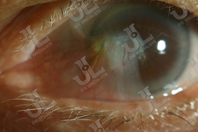
Pterygium
Pterygium is a wing-like growth over the conjunctiva and cornea of the eye. This is degenerative in nature, usually worsened by exposure to the sun or ultraviolet light (UV). As it enlarges it can obstruct the vision, causing astigmatism and discomfort from inflammation.

Managing Pterygium
When outdoors, it is best to wear UV protective eyewear and a hat or cap to decrease sun exposure. Surgical excision is recommended when the pterygium is cosmetically obvious or causes blurring of vision. The procedure involves removing the fleshy growth, as well as harvesting a conjunctival graft from the same eye and placing it over the excised area with fibrin glue. This will decrease the rate of recurrence.
Poor Ocular Surface and Limbal Stem Cell Deficiency
Poor or irregular ocular surfaces arise when there is an overuse of contact lens, or when one suffers from severe dry eyes, eyelid margin diseases or ocular surface inflammation. Patients with this condition experience severe eye discomfort, redness and poor vision.
Some cases of poor ocular surfaces are associated with limbal stem cell deficiency (LSCD), a loss or deficiency of the stem cells in the limbus that are vital for the repopulation of the corneal epithelium. When these stem cells are lost, the corneal epithelium is unable to repair and renew itself resulting in epithelial breakdown and persistent defects, scarring and inflammation.
Treating Poor Ocular Surfaces
Poor ocular surfaces can be treated with eye lubricants and anti-inflammatory medications.
For more severe cases, ocular resurfacing is done surgically through amniotic membrane transplantation. The amniotic membrane functions in the eye as a basement membrane substitute or a temporary graft. It has anti-inflammatory and anti-scarring effects and contains growth factors that promote epithelial wound healing on the surface of the eye. This tissue has been used as a tissue bandage for cornea infections and sterile melts, and to reconstruct the ocular surface for various procedures.
Limbal stem cell transplantation is done for cases associated with LSCD to treat the loss or deficiency of stem cells. Limbal stem cell grafts can be harvested from a donor and transplanted onto the ocular surface of a patient. The donor graft can be harvested from the patient’s fellow eye.
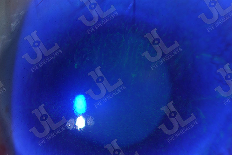
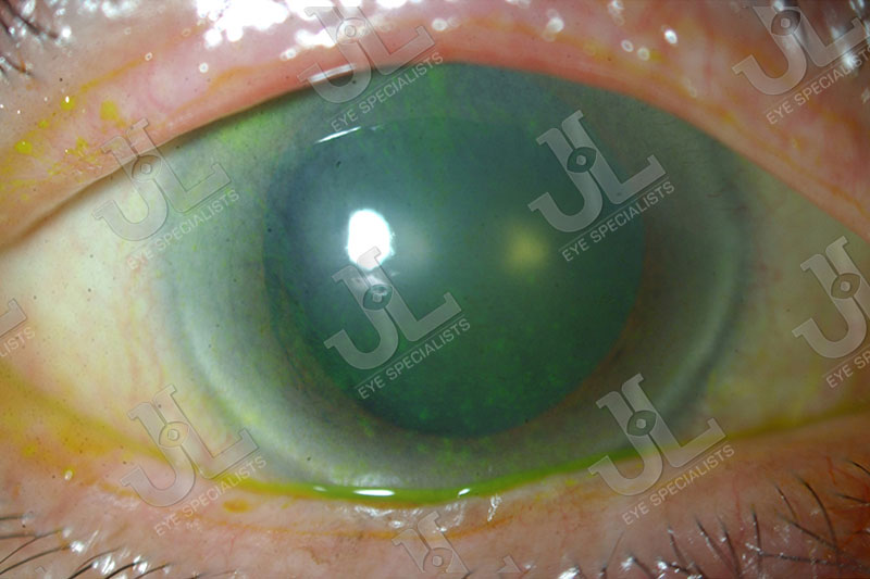
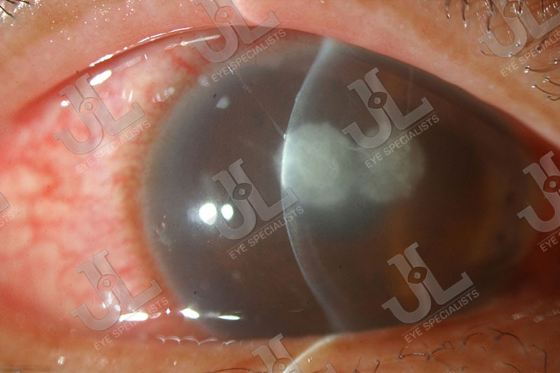
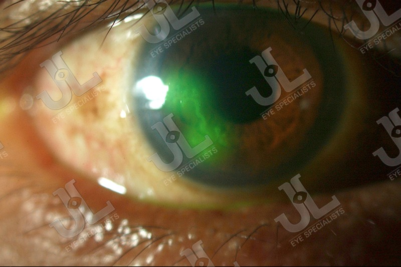
Corneal Ulcers and Corneal Infections
A corneal ulcer typically occurs as a painful infection on the surface of the eye with mild to severe eye discharge and reduced vision. In some severe cases, it can even lead to blindness.
Bacteria
Most cases of corneal ulcers are due to a bacterial infection that invades the cornea, often following injury to the eye surface. Contact lens wearers are particularly susceptible to such infections as the contact lens may rub against the eye’s surface, causing slight damage to the epithelium that enables bacteria to penetrate the eye.
Besides bacteria infection, other causes of corneal ulcers are fungus, virus or parasite:
Fungus
Fungal keratitis or keratomycosis refers to an infective process of the cornea typically caused by trauma with vegetative material. In urban society, the use of contact lens can also cause fungal keratitis.
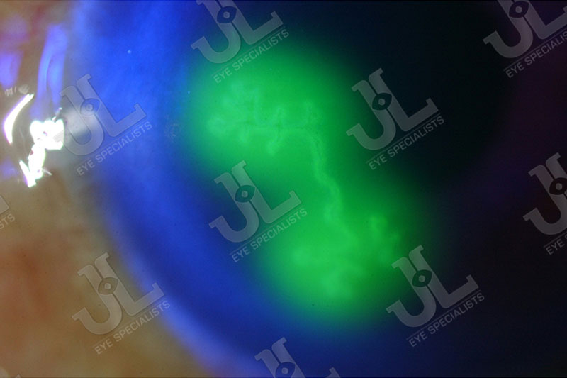
Virus
The most common, recurrent virus affecting the eye is the Herpes Simplex Virus. This type of virus can cause inflammation and scarring of the cornea that sometimes is referred to as a cold sore on the eye. Herpes of the eye can be transmitted through close contact with a person who is having an outbreak, or through self contact and contamination during an active herpes infection (such as a cold sore of the lip). The exact cause of an outbreak is unknown, but stress-related factors such as fever, sunburn, major dental or surgical procedures and trauma are often associated with it.
Parasites
Acanthamoeba are naturally occurring amoeba (one-celled organisms) commonly found in water sources, such as tap water, well water, hot tubs and sewage systems. These common parasites can enter the eye and cause acanthamoeba keratitis, a very serious eye infection that can result in permanent scarring of the cornea and vision loss.
Contact lens wearers are at increased risk of encountering acanthoamoeba infection if proper contact lens care is not followed. One should not wear contact lenses while swimming or using a hot tub.
Another common parasite is microsporidia, a spore-forming unicellular parasite found in soil. Microsporidial infection can occur when mud enters the eye during sporting activities such as football or rugby in muddy fields. This can cause keratoconjunctivitis, which may cause eye pain, redness or blurry vision as early as two days to a couple of weeks after exposure.
Managing Corneal Ulcers and Corneal Infections
Most corneal ulcers and infections can be effectively treated with intensive antibiotics, antifungal, antiviral and antiparasitic eyedrops and medication.
When infections are more severe, the treatment will be prolonged and visual outcome can be more unpredictable. There may be a need for surgical management such as a cornea biopsy to establish the diagnosis or a therapeutic cornea transplant.
Keratoconus / Keratoectasia
Keratoconus is due to progressive irregularity and distortion of the cornea from the weakness of the cornea stroma. It is an eye disease in which the regularly shaped cornea thins and begins to bulge into an irregular cone-like shape. This irregular shape results in irregular light refraction as it enters the eye on its way to the light-sensitive retina, causing distorted vision.
Keratoconus can occur in one or both eyes and often begins during the teenage years or in the early 20s. This condition causes increased irregular astigmatism and poor vision that cannot be corrected by spectacles or contact lenses. The progressive thinning of the cornea also results in scarring and discomfort of the eye.
Most causes of keratoconus are inherent weakness of the cornea stroma and worsened by rubbing of the eye. Some are due to post-refractive surgical procedures and other cornea procedures.

Management of Keratoconus
Keratoconus can be managed with rigid gas permeable contact lenses which may improve the vision but does not have any effect on its progression.
The condition can also be treated by way of Corneal Collagen Cross-Linking, which employs an innovative use of UV-B light to strengthen the corneal stroma.
For patients experiencing keratoconus or keratoectasia with severe thinning and scarring, a cornea transplant is performed to improve its clarity.
Advanced Technologies for Corneal Diagnostics and Management
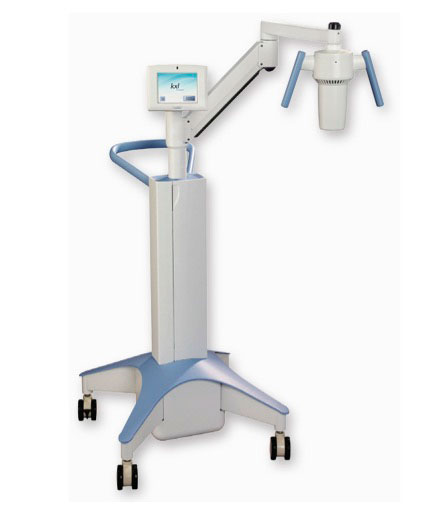
Avedro KXL
Avedro KXL system is used to perform multiple types of corneal cross-linking procedures. The system achieves accelerated cross-linking in minutes by increasing the UVA power and reducing the exposure time while maintaining the same entire energy on the eye as standard cross-linking.
Also, the KXL system is the first and only system manufactured with the highest power available, superior top hat beam profile, detailed depth of focus, wireless control for beam alignment, fully integrated stable delivery platform, complete with a touch screen monitor for programming and controlling of the machine.
The KXL system’s higher power yields shorter UVA exposure times for increased patient safety, comfort and improved practice efficiency.

Nidek Specular Microscope
The Nidek specular microscope measures the paracentral corneal cells comprehensively. Its two-seconds analysis measures the endothelial cells from the quantity to the endothelial cell density. The advanced manual analysis function allows the clinician to select the cells manually and ease the process of forming a diagnosis.
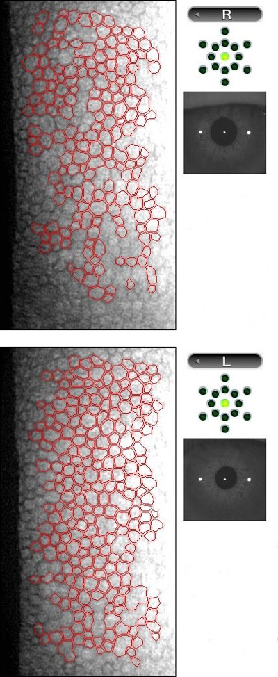
Image 1.1
Image 1.1 is a report of a patient who has a normal endothelial cell quality where the results show a healthy cell count, cell density, area, shape of cells, and corneal thickness.
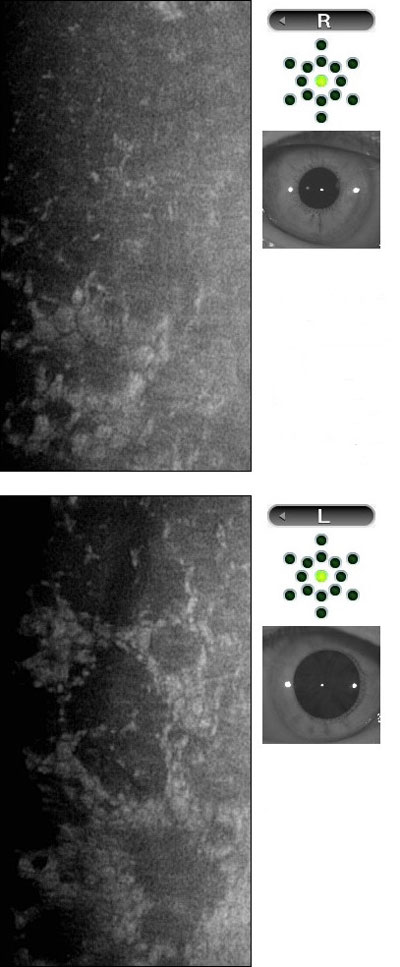
Image 1.2
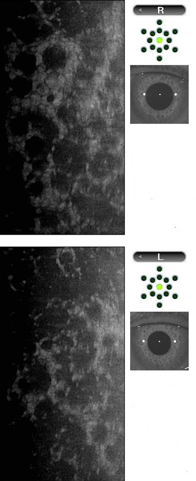
Image 1.3
Images 1.2 & 1.3 show a patient with Fuch’s endothelial cell dystrophy, a condition where the cornea becomes edematous as cells are lost over time.
Galilei G4
The Galilei G4 combines Placido disc–based topography and Dual-Scheimpflug tomography in one unit. This combination allows the device to provide complete analysis of both the anterior and posterior corneal surface for comprehensive screening required for refractive surgery such as LASIK, PRK, ASA, etc. all in one measurement session.
The Galilei G4 topography function accurately measures the anterior curvature of the cornea where it precisely detects anterior surface irregularities and tear film quality.
The Galilei G4 tomography function precisely measures pachymetry and elevation data with 3D anterior chamber analysis. The ray-traced posterior corneal surface data detects any protrusion or asymmetry in early stages of corneal diseases.
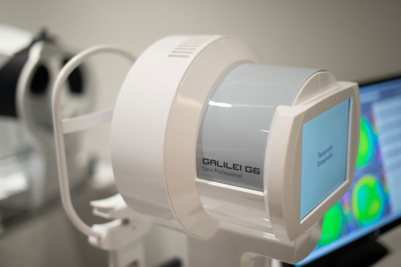

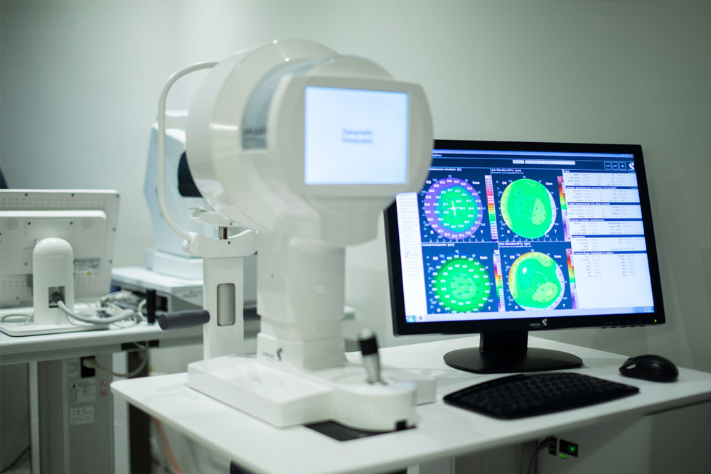
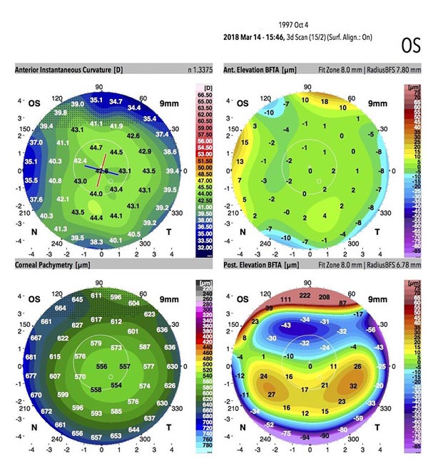
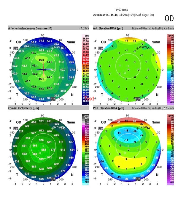
Image 2.1
Image 2.1 illustrates the topographical scan of the cornea that is normal. As seen, the scan shows a Keratoconus Prediction Index (KPI) shows a value of 0.0%. From the scan, it is evident that the cornea is even and regular. The scan also does a scan of the thickness of the cornea where normal count of an average person is around 540 microns. Image 2.1 shows a corneal thickness of 551 to 651 microns from the thinnest section to the periphery of the cornea and a central corneal thickness of 556 microns, which indicates a healthy range.
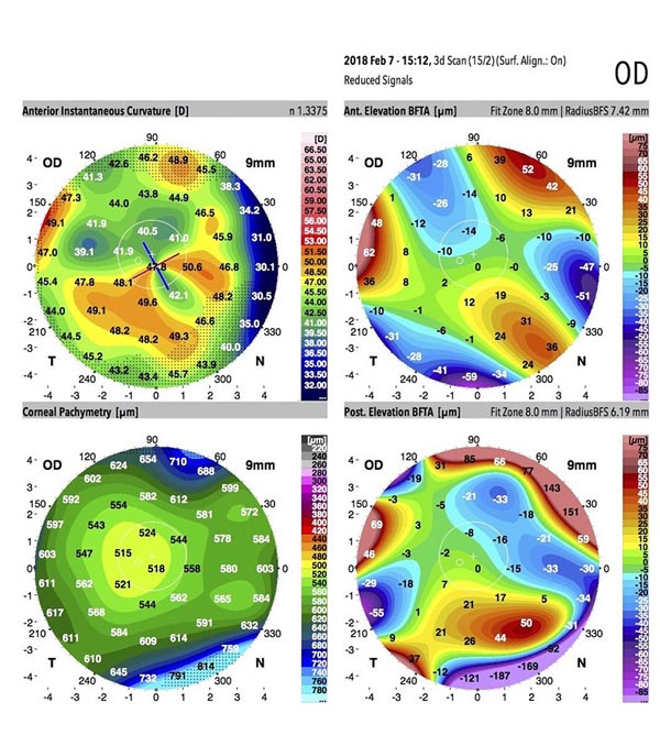
Image 2.2
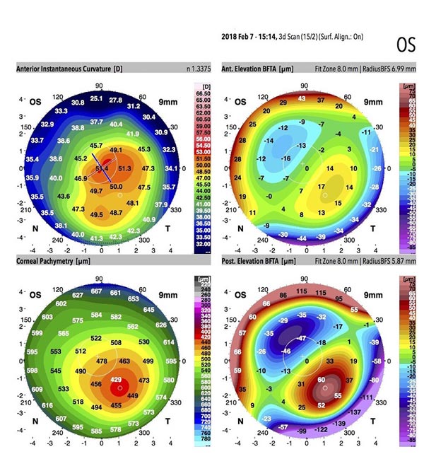
Image 2.3
Images 2.2 & 2.3 shows a report of a pair of abnormal corneas of a person. Image 2.2 KPI shows an index of 32.7% and image 2.3 shows a KPI of 93.8%. The corneal thickness of image 2.2 is from 512 to 660 where the corneal thickness is only 518 microns.
Whereas, for image 2.3, the corneal thickness is from 422 to 622 microns and the central corneal thickness is at 478 microns. The central corneal thickness is important for keratoconus as the definition describes it as a progressive thinning of the central cornea that causes the cornea to appear cone-like. Therefore, by performing such accurate measurements swiftly, we are able to detect the abnormalities of the eye.
Book An Appointment With Us
We understand that each patient comes with their unique concerns. We customize our approach to ensure that we provide each patient with the appropriate treatment to address their needs.